Brain Tumor Detection with Deep Learning
Leveraging AI to assist in the early and accurate identification of brain tumors from MRI scans.
Python · TensorFlow/Keras · Deep Learning · Medical Imaging
Project Significance
Early detection of brain tumors is critical for patient outcomes, but interpreting MRI scans is a complex, time-consuming task prone to human error. My motivation was to develop an AI-powered diagnostic aid: a fast, accurate, and reliable tool to provide clinicians with a crucial "second opinion," especially valuable in regions with limited access to specialized medical expertise.
Solution Developed
I engineered and trained a sophisticated deep learning model specifically for the multi-class classification of brain tumors from MRI images. The dataset comprised over 7,000 brain scans, encompassing various tumor types (glioma, meningioma, pituitary) and healthy controls. The primary objective was to achieve high discriminative accuracy in differentiating between these categories.
Methodology & Approach
I managed the entire project lifecycle, from initial data acquisition and rigorous preprocessing to model training and robust evaluation. This involved:
- Data Engineering: Identified, cleaned, and augmented the MRI dataset to ensure sufficient variability and reduce bias, crucial for model generalization.
- Model Architecture: Designed and implemented a custom Convolutional Neural Network (CNN) architecture using Python with TensorFlow/Keras, optimizing for image feature extraction and classification.
- Training & Evaluation: Monitored training progress, loss functions, and accuracy metrics. Performed thorough validation using standard metrics like accuracy, precision, recall, and F1-score, visualized through confusion matrices to ensure robust performance across all classes.
- Interpretability (Implicit): While not explicitly detailed, the focus on accuracy and clear evaluation metrics implies an understanding of model performance critical for medical applications.
Key Results & Learnings
- Achieved a **test accuracy of 99%** on unseen MRI scans, demonstrating exceptional performance in differentiating between tumor types and healthy brains.
- Developed a solution capable of providing fast and consistent predictions, offering significant potential to assist medical professionals, particularly in resource-constrained environments.
- Gained profound insights into the unique challenges and ethical considerations of applying deep learning in healthcare, reinforcing the importance of rigorous testing and validation.
Learnings & Reflections
This project transcended pure technical execution; it was a deeply rewarding endeavor with tangible humanitarian potential. It instilled in me greater confidence in my ability to independently navigate complex data science challenges from inception to deployment. The experience underscored the immense power of data and AI to drive meaningful societal change and improve human lives, solidifying my passion for applying data science to real-world impact.
Full Tech Stack Used
- Languages: Python
- Libraries: TensorFlow, Keras, NumPy, Pandas, Matplotlib, OpenCV
- Tools: Jupyter Notebook, Google Colab
- Concepts: Deep Learning (CNNs), Image Classification, Data Augmentation, Model Evaluation
Visual Highlights
A typical MRI image from my dataset, used for training and prediction.
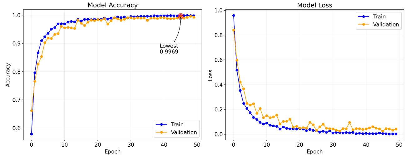
Visualization of the model's accuracy improvement during training over epochs.
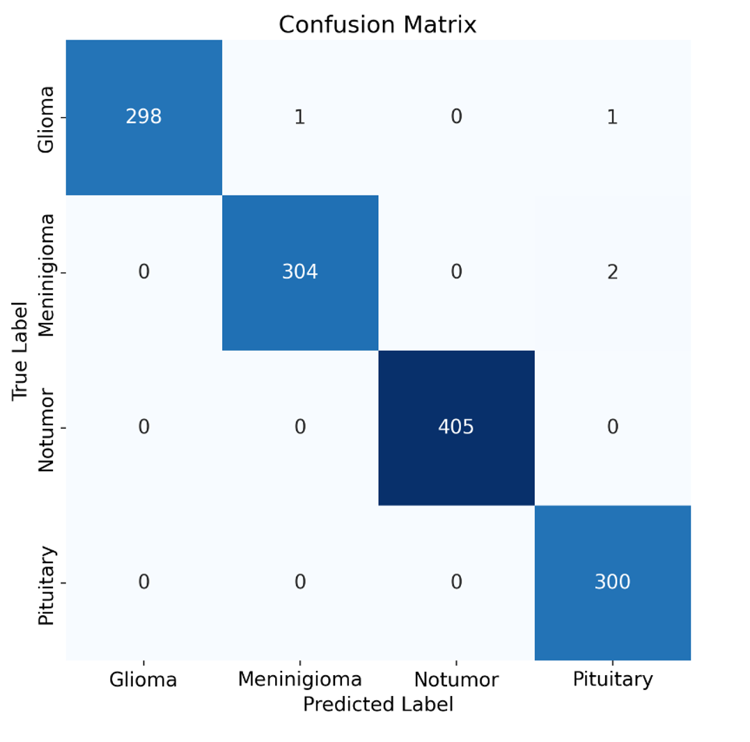
A confusion matrix detailing the model's classification performance across different tumor types and healthy scans.
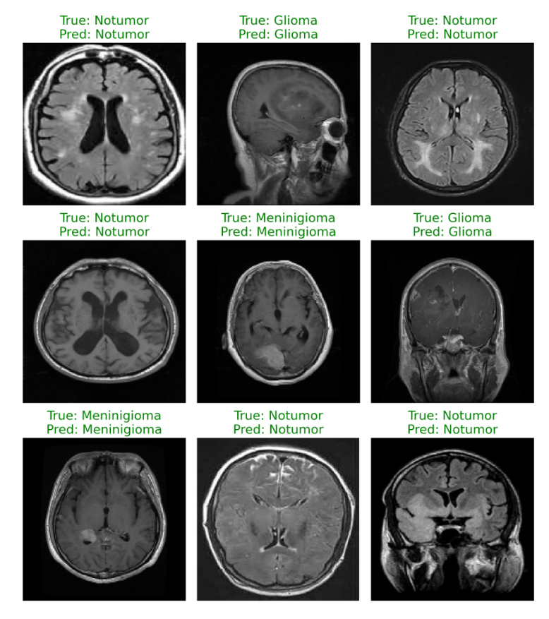
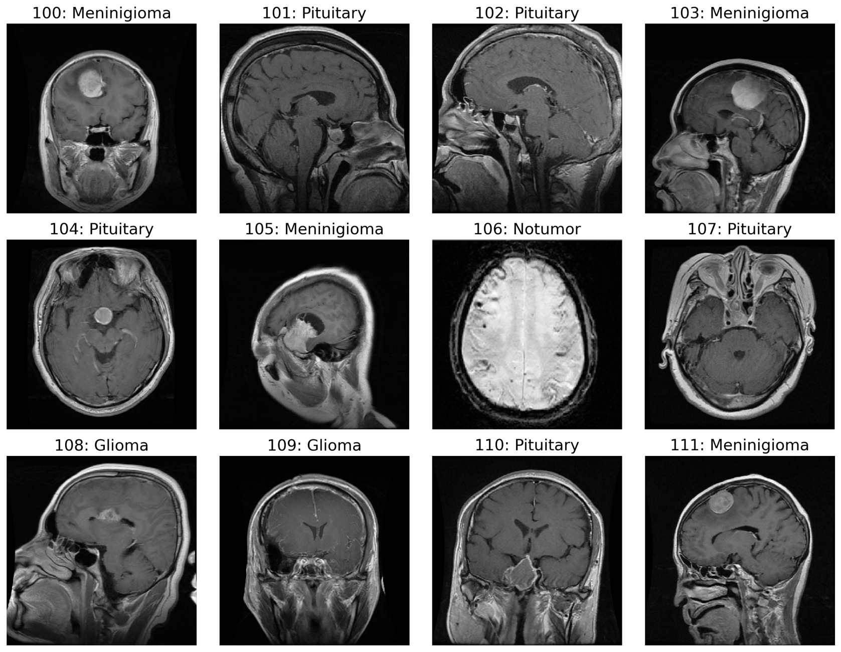
Examples of the model’s predictions on unseen MRI images from the test set.